
AP, Mortise and lateral view of the right ankle in case 2. Yellow... Download Scientific Diagram
Ankle Fracture Mechanism and Radiography. Robin Smithuis. Radiology Department of the Rijnland Hospital, Leiderdorp, the Netherlands. The ankle is the most frequently injured joint. Management decisions are based on the interpretation of the AP and lateral X-rays. In this article we will focus on:
Assessing Heel Pain Diagnostic Ultrasound of the Foot and Ankle
The x-ray beam is then angled cephalad about 40 degrees to the long axis of the foot and first penetrates the sole of the foot. The dorsoplantar view is performed with the patient prone with a wedge or pillow placed under the distal calf.. In the AP ankle view with passive hindfoot varus stress 12 degrees of talar tilt or 5 degrees more than.

Xray Image of Ankle, AP and Lateral View. Stock Image Image of joint, metatarsal 53839873
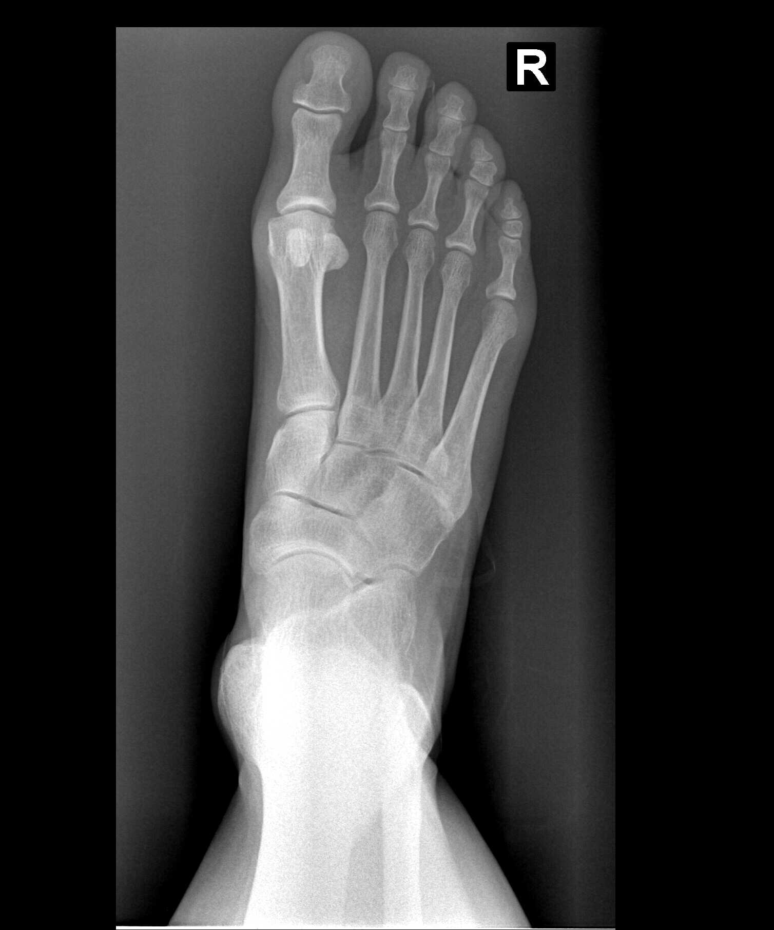
AP images are obtained by directing the x-ray beam from the dorsum of the ankle to the plantar surface, with the image receptor beneath the sole of the foot. Internal oblique images are obtained by internally rotating the ankle 15-20 degrees and directing the x-ray beam in a dorsoplantar direction similar to the AP view.
The Ankle
An isolated fracture of the medial malleolus, or widening of the ankle joint with no visible fracture seen on ankle X-ray, should raise the suspicion of an associated fracture of the fibula. If this is not visible in the distal fibula then further X-rays of the proximal fibula should be performed. Imaging of the proximal fibula should also be.

Pin on ankle joint ap view basic Anatomy
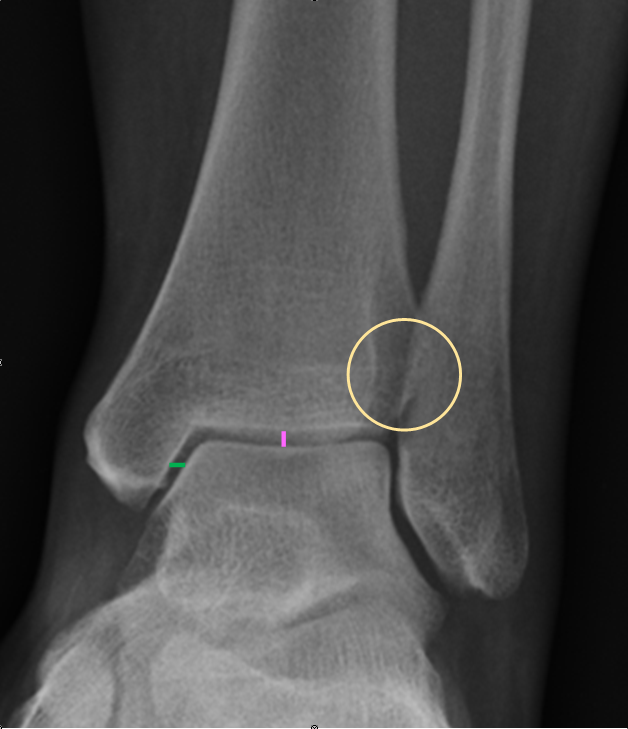
Interpret traumatic ankle x-rays using a standard approach; Identify clinical scenarios in which an additional view might improve pathology diagnosis; Why the ankle matters and the radiology rule of 2's The Ankle.. Tibiofibular clear space: On AP view, this is the distance between the medial border of the fibula and lateral border of the.

Normal values at standard Xray views (AP,Mortise and lateral)of the... Download Scientific
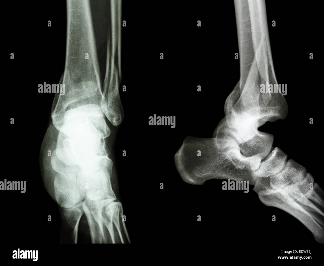
Ankle views. An x-ray of the ankle will have three views - AP, mortise, and lateral. It should be noted, though, that in some countries, including the UK, only the mortise and lateral are used. See the annotated images below from WikiFoundry, and thanks also to Radiopaedia:
Rt AP Foot Xray 1012013
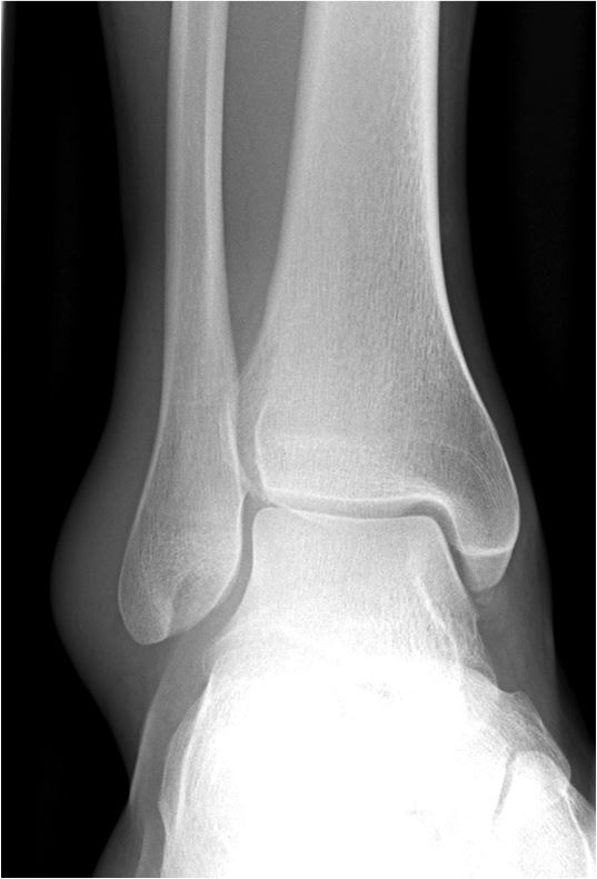
same horizontal plane as the medial malleolus and both are parallel to the x-ray tabletop. The mortise view is the true AP projection of the ankle joint. Oblique projections, 1 plain radiograph tomography , computed tomography (CT), or magnetic resonance imaging (MRI) may be required to identify minimally displaced ankle fractures.
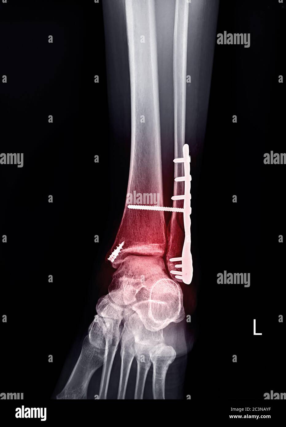
film xray ankle AP/Lateral show fracture distal tibia and fibula (leg's bone) and ankle joint
This video tutorial presents the anatomy of ankle x-rays:0:00. Intro to ankle x-rays0:13. Standard ankle series for x-rays0:20. AP view (right ankle)1:27. Mo.
Normal ankle joint, Xray Stock Photo Alamy
Citation, DOI, disclosures and article data. The ankle series is comprised of an anteroposterior (AP), mortise and lateral radiograph. The series is often used in emergency departments to evaluate the distal tibia, distal fibula, and the talus; forming the ankle joint. See approach to an ankle series.
Ankle Xray Interpretation Ankle Fracture Geeky Medics
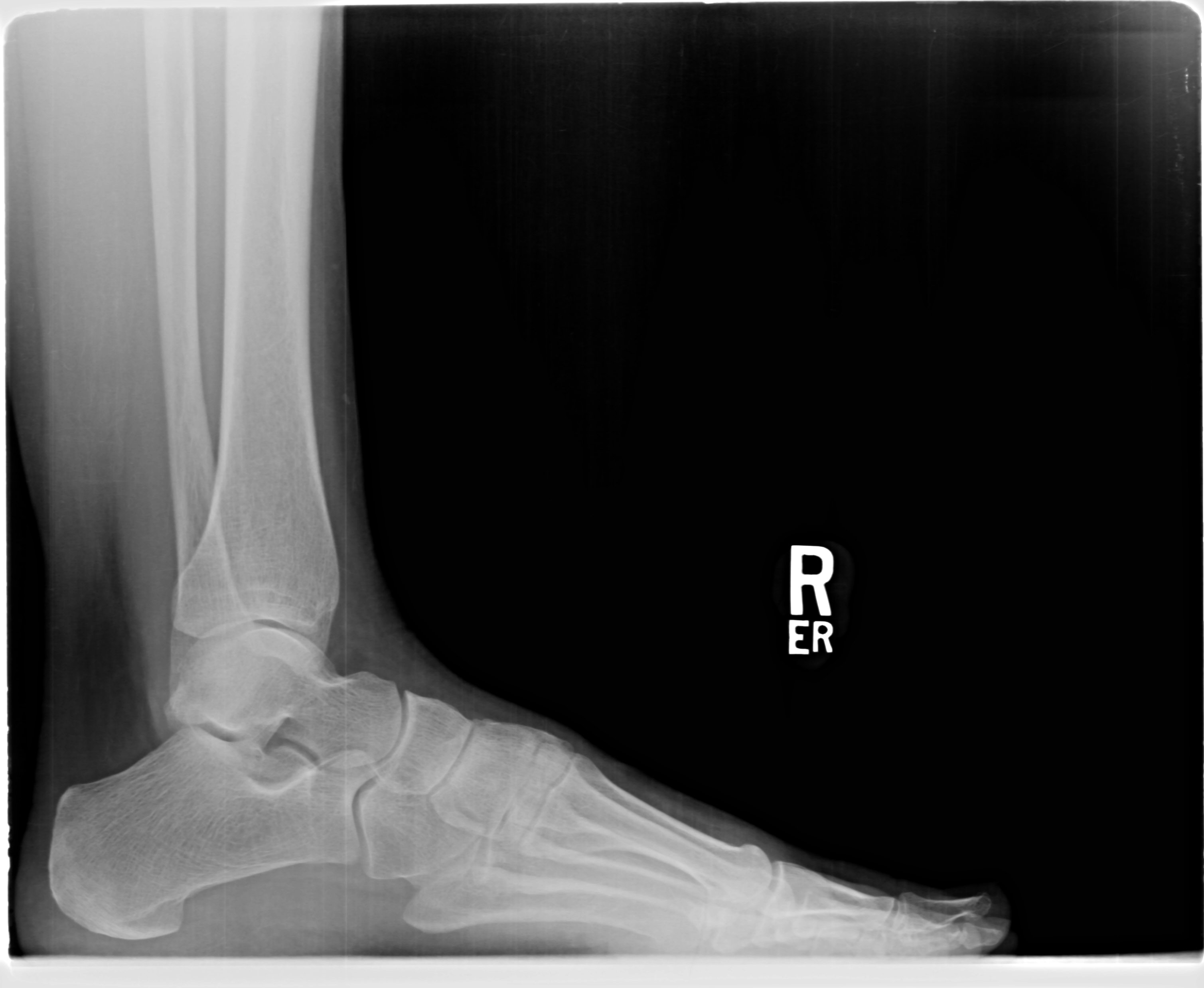
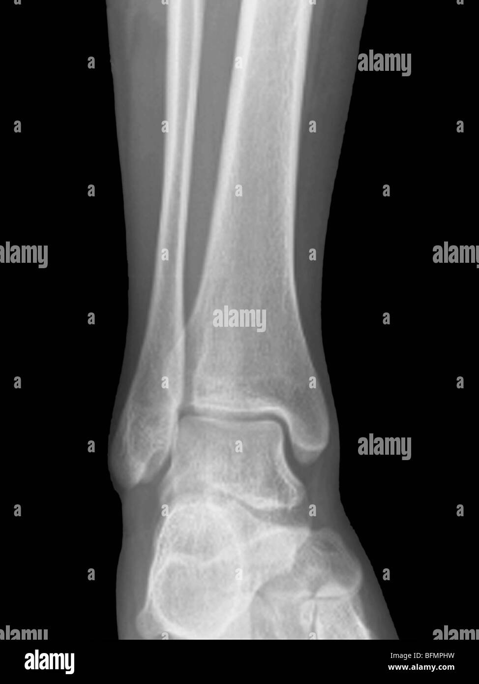
An ankle x-ray, also known as ankle series or ankle radiograph, is a set of two x-rays of the ankle joint. It is performed to look for evidence of injury (or pathology) affecting the ankle, often after trauma.. AP and lateral views of the ankle. AP view performed at a slight angle to open up the mortise; similar tests. tib/fib x-ray.

AP and lateral radiograph of the left ankle showing ball and socket... Download Scientific Diagram
This projection is utilized to assess the structural integrity of the ankle joint. If the patient is able, weight-bearing views should be performed in acute and follow up settings 1 . In addition, this view can show bony diseases or lesions of the distal lower leg, talus and proximal fifth metatarsal. Ultimately the radiographer will determine.

EMRad Radiologic Approach to the Traumatic Ankle
- on AP view differnece in width of superior clear space between medial and lateral side of the joint should be < 2 mm; - these are static measurements of the talar position; - in normal ankle, talus may tilt up to 5 deg w/ inversion stress; - measurements of talar tilt using stress x-rays are used to evaluate lateral ligament stability.
Xray ankle or Radiographic image or xray image of left ankle joint AP view showing ankle plate
A structured approach to ankle X-ray interpretation to identify fractures and other abnormalities. The guide includes X-ray examples of key pathology.. Mortise view: this is a modified anteroposterior (AP) view of the ankle in 10-20° internal rotation so that the medial and lateral malleoli are in the same horizontal plane and joint.

Ankle X Ray Anatomy
Stress view. Positioning. patient. manual stress = supine + knee extended + ankle inverted/everted. gravity stress = supine + hip ER + knee flexed + ankle placed on bump. beam. aim at tibiotalar joint. Uses. joint stability = < 5° difference between ipsilateral + contralateral ankles.

Lateral and anteroposterior (AP) stress ankle radiographs... Download Scientific Diagram
On a true AP-view the talus overlaps a portion of the lateral malleolus, obscuring the lateral aspect of the ankle joint.. This was the only fracture that was seen on the x-rays of the ankle and this patient turned out to have an unstable Weber-C fracture and went for surgery. The x-ray beam has to be centered on the malleoli.

Xray picture of the right ankle joint in the AP view. Thick ening... Download Scientific Diagram
Central ray 10 degrees cephalad at a point 1 inch (2.5 cm) distal to the medial malleolus. LE-P-30 - Ankle AP. Purpose and Structures Shown AP projection of ankle joint, distal ends of tibia and fibula, and proximal portion of talus. Position of patient Supine position. Affected limb fully extended.